Below you can view pictures of Morgellons Disease:

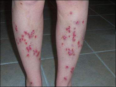

Image sequence 1 of Morgellons patient. Note the ink outline
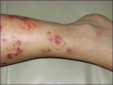
Image sequence 2 of Morgellons patient. Closer view of outline area.
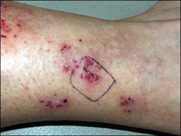
Image sequence 3 of Morgellons patient. Zooming in on outline area.
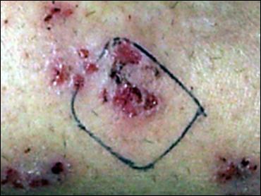
Image sequence 4 of Morgellons patient. Outline area magnified 2x.
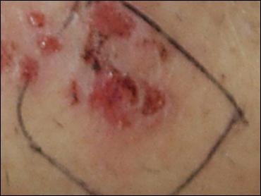
Image sequence 5 of Morgellons patient. Outline area magnified 4x.
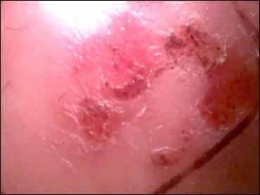
Image sequence 6 of Morgellons patient. Outline area magnified 10x – note ink line at right.
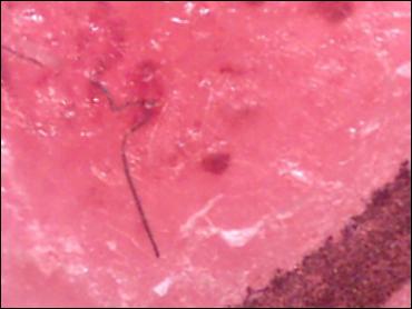
Image sequence 7 of Morgellons patient. Outline area magnified 60x – note ink line still present.

Image sequence 8 of Morgellons patient. Outline area magnified 60x.
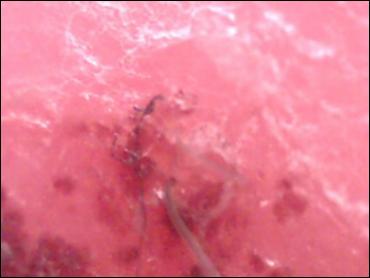
Image sequence 9 of Morgellons patient. Outline area magnified 60x – note hair in foreground.
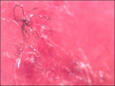
Image sequence 10 of Morgellons patient. Outline area magnified 80x.
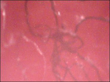
Image sequence 11 of Morgellons patient. Outline area magnified 200x.

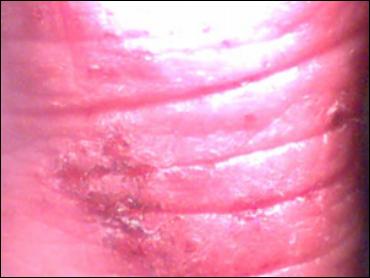
Lip of 3-year-old male at 10x showing fibers embedded in skin.
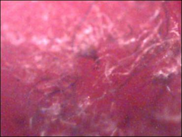
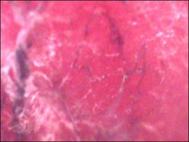
Fibers removed from same lip lesions shown at 60x.
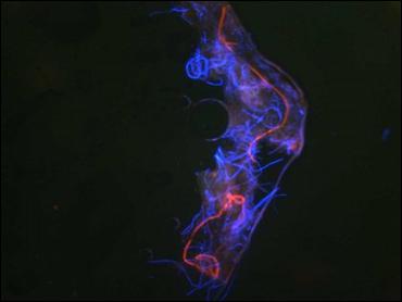
Image showing autofluorescence of fibers from child’s lip skin lesion.
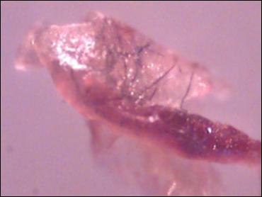
Heel of 3-year-old male at 10x showing lesions with associated fibers.
Same heel lesions on 3-year-old male showing fibers at 60x.
Same heel lesions on 3-year-old male showing fibers at 60x.
User Submitted Pictures
Have a picture to share relating to Morgellons Disease? Please click here to upload one.


